Autonomous Software-Driven Microscopy Focusing System #
Abstract #
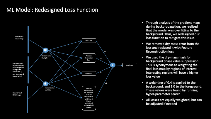
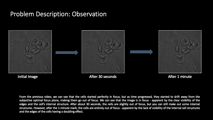
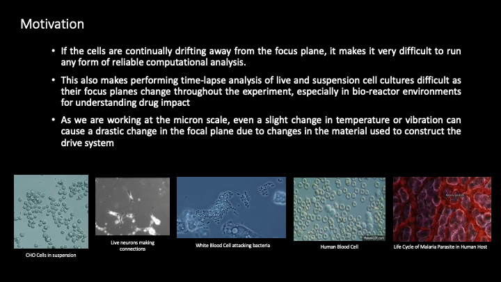
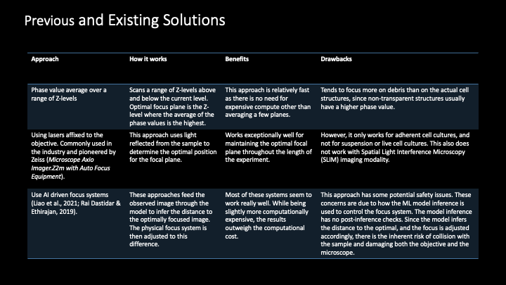
Focus drift presents a substantial challenge in time-lapse microscopy imaging, especially during extended imaging sessions of live and suspension cell cultures in bioreactor environments. Existing methods, such as hardware-based adjustments or software solutions that rely on lasers and average phase value computations, often fall short by either being limited to specific types of cell cultures or by focusing on the debris rather than the sample. Recent advances include AI-driven solutions that directly adjust the focus based on the current observation in an end-to-end fashion. These systems work exceptionally well compared to pure hardware or software-based solutions. However, they lack safety measures and risk damage to the samples and to the microscope through objective-sample collision.
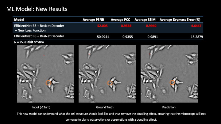
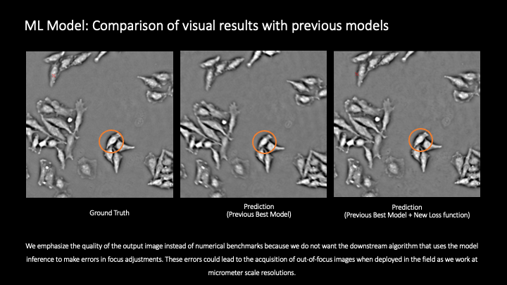
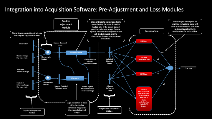
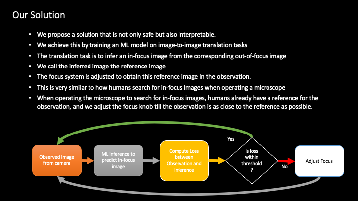
To address these challenges, we develop the Autonomous Software-Driven Microscopy Focusing System. This system introduces a machine learning based solution that significantly enhances focus stability and accuracy. Our novel approach utilizes an image-to-image translation task, where the model predicts in-focus images from the corresponding out-of-focus images. This predictive capability is then used to actively adjust the microscope’s focus, ensuring the observation closely matches the reference image, thereby mimicking the natural focusing method used by humans.
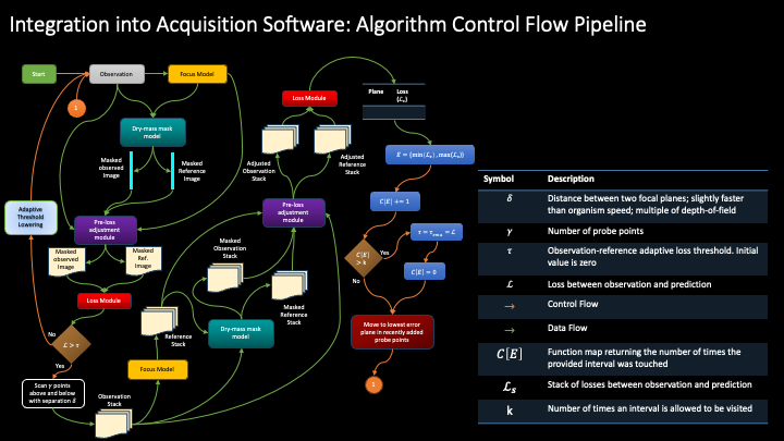
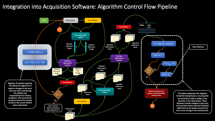
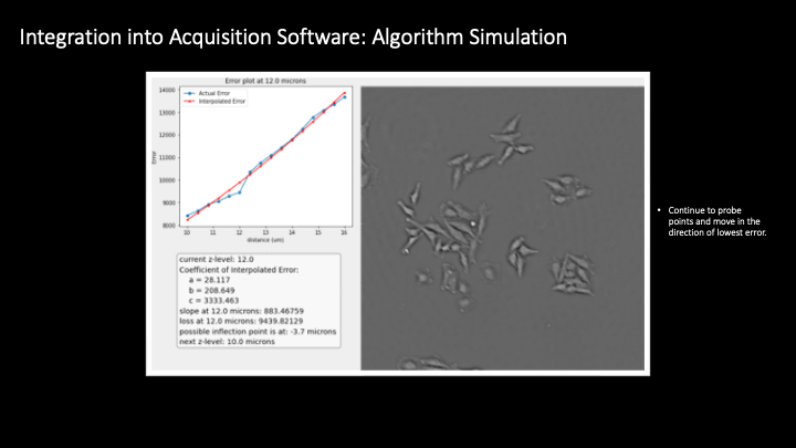
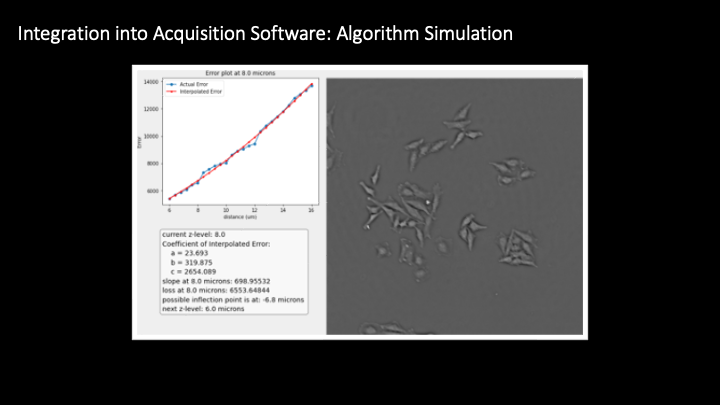
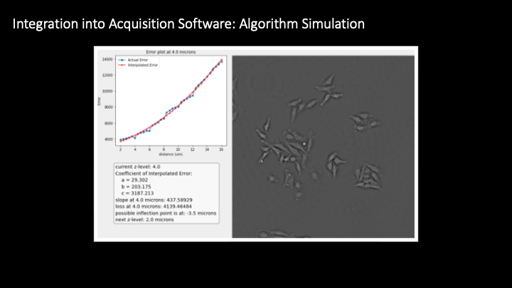
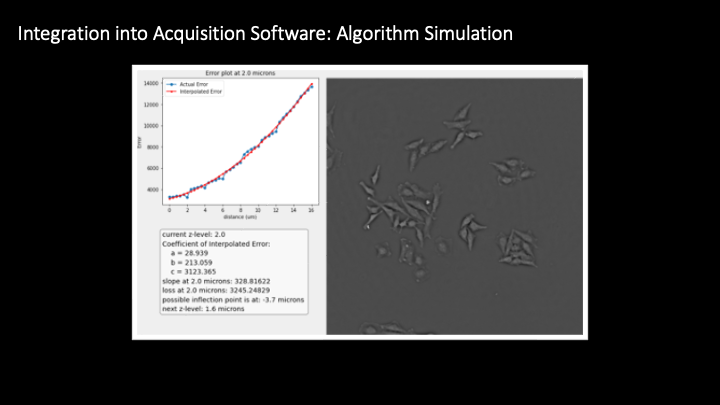
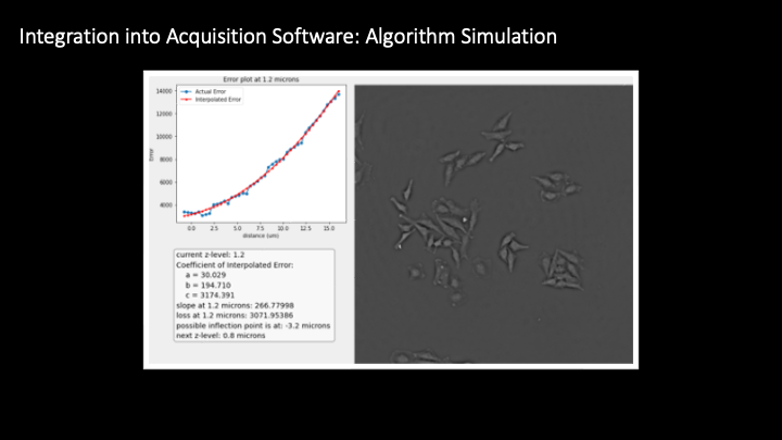
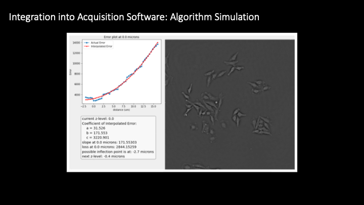
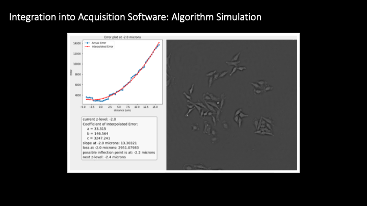
Furthermore, to ensure inherent safety associated with our system, we pass the model inference through a post-inference pipeline algorithm that is responsible for making adjustments to the focus drive of the microscope.
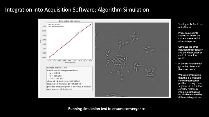
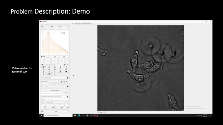
Our system is fine-tuned to handle dynamic changes in the imaging field without compromising sample safety. This method not only prevents focus drift but also supports the acquisition of high-quality images critical for reliable computational analysis. Demonstrated in real-time application in the CellVista acquisition software, this approach proves to be a robust solution, combining safety and interpretability, thus marking a significant advancement in biomedical imaging techniques.
Presentation #